Neurones are beautiful and fascinating. Scientists make an artform of recording their research. Click below to see some incredible images of neurones. (AS revision - what kind of microscopes and techniques have been used to make these images?).
To begin, let's look at an example of how a stimulus in the external environment of an organism is transduced into an electrical impulse along a nerve fibre to the brain.
Click on the images below to look at an animation showing how vibrations are interpreted as sounds by our brain (EXTN) and how skin receptors detect changes in our external environment.
Neurons
Neuron Structure in CNS: Neurons are the specialized cells that control and monitor body activities and physiological functions. They sense changing conditions, process sensory input, and direct the body’s responses. Click here to see a short animation introducing the structure of a neuron.
Synoptic extension - a video about Multiple Sclerosis.
Multiple Sclerosis: The central nervous system comprises nerve cells, or neurons, in the brain and spinal cord. A fatty product called myelin surrounds most nerves in the body. Myelin allows the nerves to send clear electrical impulses faster and more effectively along the neurons. Multiple sclerosis (MS) is an autoimmune disorder, which means that the body’s immune system turns against itself. This autoimmune response causes the protective myelin coating, or myelin sheath, to become inflamed and eventually destroyed in various places along the central nervous system. This destruction of myelin is called demyelination. This destructive process keeps the neurons from sending effective nerve signals. The signals become slowed, garbled, or blocked, causing the symptoms of multiple sclerosis to develop. Symptoms of MS are varied and depend on the location of the myelin damage. Common symptoms are loss of muscle coordination, impaired vision, numbness or tingling sensations in the arms or legs, fatigue, and incontinence. This disease can be difficult to diagnose because the symptoms, which can last from days to months, may come and go without any pattern.
The reflex arc (GCSE REVISION)
The Resting Potential
Click on the image below to see how a neuron maintains a resting potential.
The resting potential is maintained due to the active transport of Potassium and Sodium ions across the plasma membrane. The protein responsible for this is the sodium-potassium pump.
QUIZ Click below for a good tutorial on how this pump works and a quiz to reassure yourself that you understand the process correctly.
The Action Potential
Click here to see an animation of an Action Potential moving down an axon.
QUIZ
EXTN: How do Scientists measure these tiny changes in current!? By using a really large axon - the giant axon of a squid.
Here is a BRILLIANT video showing how scientists in the 1970s used the squid axon to make many of the discoveries you have learnt so far in your studies.
Revise and test yourself on the entire process by following through the tutorial link below.
Here are four more quizzes on the action potential to test your overall understanding.
QUIZ 1 on the role of voltage gated channels in the action potential
QUIZ 2 on the role of voltage gated channels in the action potential
QUIZ 3 : an action potential on an unmyelinated axon.
QUIZ 4 : an action potential on an unmyelinated axon.
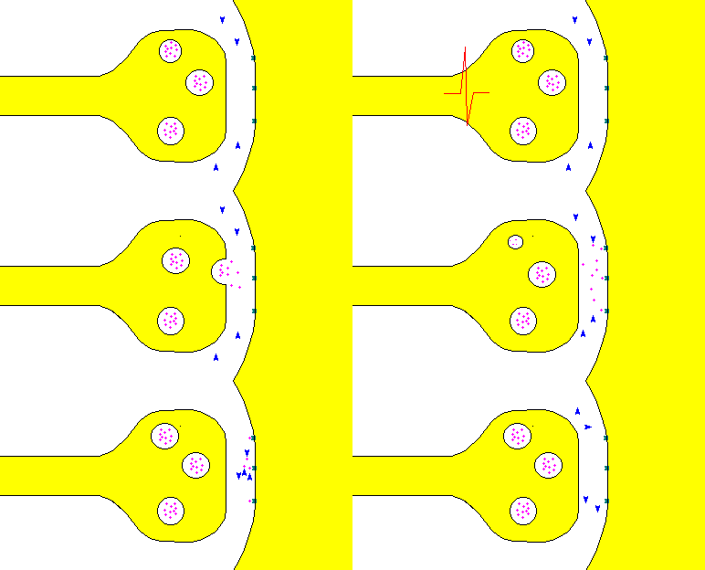
Saltatory conduction
Continuous and Saltatory Propagation: When an action potential in an axon spreads to a neighboring region of its membrane by a series of small steps, the process is called continuous propagation. Click on this link to see a video.
24. Candidates should be able to draw a labelled diagram of a synapse; synaptic knob,
synaptic vesicle, mitochondria, pre-synaptic membrane, synaptic cleft, post-synaptic
membrane.
25. Transmission of the impulse across the
synapse is chemical rather than electrical.
The chemical, or neurotransmitter, is
enclosed in synaptic vesicles at the end of the
axon before the synapse.
The arrival of a
nerve impulse depolarises the presynaptic membrane of this axon and calcium ions rush
in. This influx of calcium causes the vesicles to fuse with the presynaptic
membrane and releases their contents into the synaptic cleft.
The neurotransmitter diffuses
across the cleft and depolarises the postsynaptic membrane, propagating the
impulse along the axon of the adjacent nerve cell.
SYNOPTIC -> Click below for an animation including how many of the cellular structures (organelles and cytoskeleton) of the presynaptic neuron are involved in the overall process.
QUIZ:
Types of Neurotransmitters
Acetylcholine and the cholinergic synapse.
Chemical Synapse: Cholinergic Synapse: A neuron relays information to another neuron at a synapse. The neuron transmitting the information is called the pre-synaptic neuron, and the neuron receiving the information is the postsynaptic neuron. Let’s look at what happens at a cholinergic synapse.
Chemical Synapse: Cholinergic Synapse at Neuromuscular Junction: A cholinergic synapse is specialized cell-to-cell junction where nerve impulses are transmitted via the release of the neurotransmitter, acetylcholine, from one neuron to another neuron or to a non-neuronal cell, such as a skeletal muscle cell.
26. The most common transmitter substances are acetyl choline and noradrenaline.
EXTN: How do insecticides work?. Describe the
effects of chemicals on synaptic transmission as illustrated by
organophosphorus insecticides acting as cholinesterase inhibitors.
Summation
Quick quiz - which letter above represents an inhibitory post synaptic potential?
This first youtube clip starts a bit slowly but bear with it because he explains simple summation nicely.
EXTN: I love this next clip - but it's really quiet! Its great at explaining how excitatory and inhibitory post synaptic potentials work together for a neuron to fire - or not!
Psychoactive drugs such as cocaine and cannabis affect synapses in the brain.
Click here to find out how different drugs disrupt the synapse and what the consequence is.


No comments:
Post a Comment