A wacky history of The Cell Theory
Can you remember the difference between plant and animal cells (GCSE!)
click here to find out.
How do we see cells?
Light microscopes
The original microscopes used light and lenses to magnify whatever was on the stage. The light microscopes we use at school work the same way.


Below are a few videos showing how Light microscopes work.
 |
| A photomicrograph is only able to have one plane of focus. |
Optical Microscopes - uses wavelengths of between 400 - 700nm. You cannot distinguish between structures closer than 200nm using a light microscope. They will appear as one object. But the light microscope cannot produce higher magnification without losing much resolution.
Laser scanning microscopes (aka confocal microscopes)
Laser scanning confocal microscopes use laser light to take many photos at different layers of the sample. The unfocused light from other planes is blocked out. If you take many in focus photos and combine them together, you are able to get a 3d image. They are usually stained - sometimes with florescence - so you can identify where in a cell certain structures are.

Laser (confocal) microscopes - have high resolution and high contrast. You can select which depth you would like to look at, you can focus on different structures at different depths in the specimen.
You are able to observe living specimens so can be used for medical uses such as looking at fungal growth in parts of the body.
The pictures below show the same specimens with traditional light microscopy (top line) and the confocal microscopy (bottom line).


Electron Microscopes.
Electron microscopes have been developed to provide images of a much higher resolution. They use a beam of fast travelling electrons with a wavelength of 0.004 nm. So they have a greater resolution than the optical microscope.

Transmission electron microscopes
- An electron beam pass through the specimen. The electrons that pass through the specimen are focused on a screen or photographic plate.
- The specimen is chemically fixed (dehydrated and stained with metal salts) so are dead.
- Produces a 2D black and white image (an electron micrograph)
- can magnify up to 2 million times.
Scanning electron microscopes
- Electrons are fired onto a whole specimen and electrons from the specimens surface bounce off and are focused on a screen.
- Specimen can be whole but is viewed in a vacuum so is dead. Specimens are also coated in metal salt stains.
- Image is 3D and black and white (can be coloured using computers).
- Magnification of x15 to x200 000.
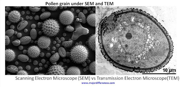
 |
| A: SEM B: TEM |
Electron microscopes
- are large and expensive.
- need a great deal of skill while using them.
- specimens are dead.
- metallic salts used to stain the specimens could be hazardous to the user.
Calculating Magnification
When using a microscope it is important that we know how big the specimen we are looking at is.
We do that by calculating magnification.
We do that by calculating magnification.
We can even measure specimens size using a stage micrometer.


No comments:
Post a Comment